

ALBUQUERQUE, N.M. — When German physicist Wilhelm Röntgen discovered X-rays in the late 1800s while experimenting with cathode ray tubes, it was a breakthrough that transformed science and medicine. So much so that the basic concept remains in use today. But a team of researchers at Sandia National Laboratories believes they’ve found a better way, harnessing different metals and the colors of light they emit.
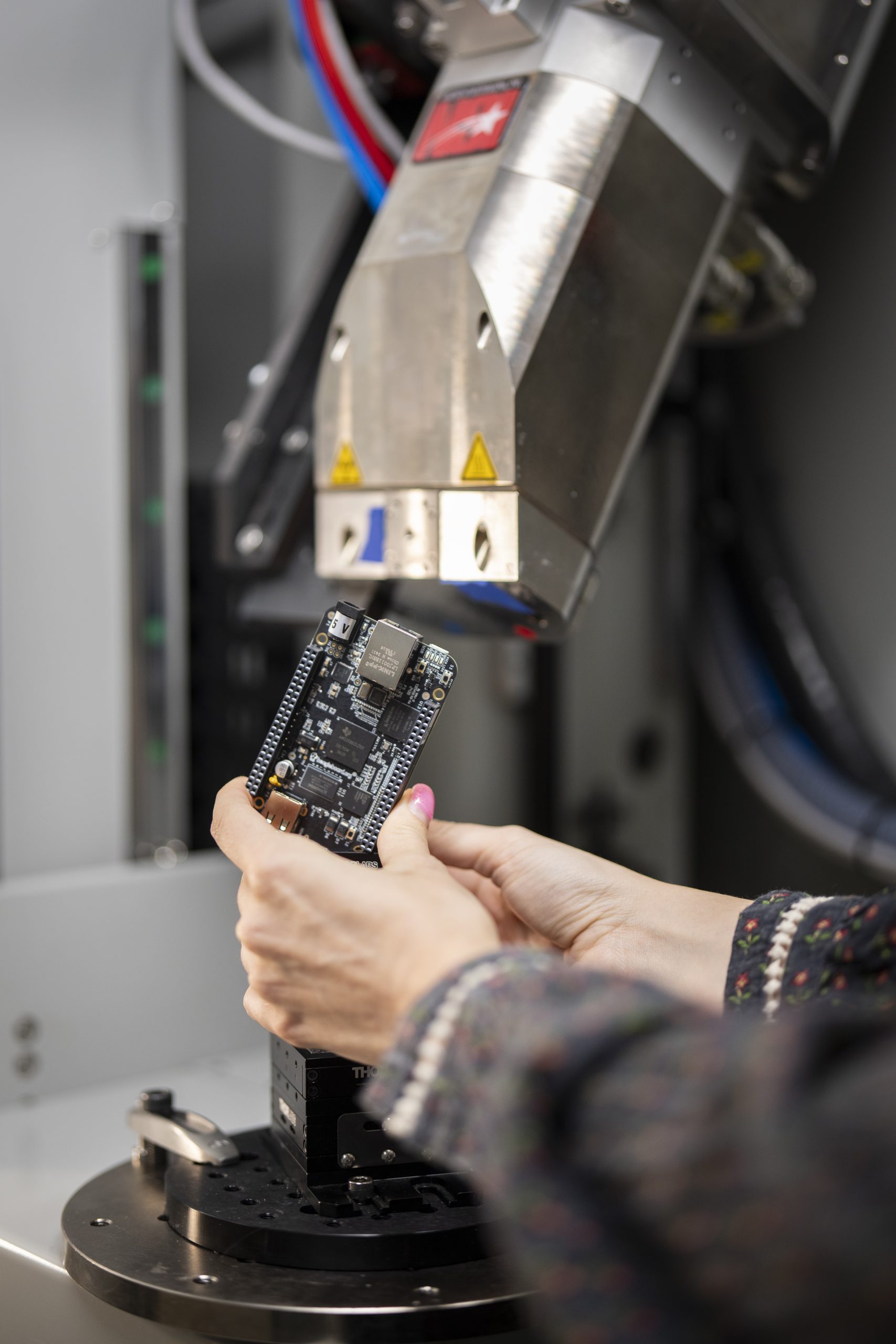
“It’s called colorized hyperspectral X-ray imaging with multi-metal targets, or CHXI MMT for short,” said project lead Edward Jimenez, an optical engineer. Jimenez has been working with materials scientist Noelle Collins and electronics engineer Courtney Sovinec to create X-rays of the future.
“With this new technology, we are essentially going from the old way, which is black and white, to a whole new colored world where we can better identify materials and defects of interest,” Collins said.
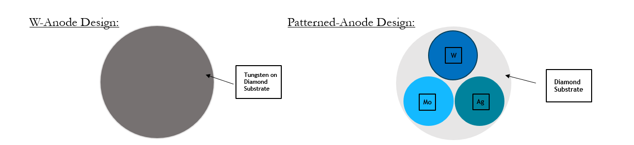
The team found they could achieve this using tiny, patterned samples of varied metals such as tungsten, molybdenum, gold, samarium and silver.
The Basics of X-ray Creation
To understand the concept, one must understand the basics of X-ray creation. Traditional X-rays are generated by bombarding a single metal target, or anode, with high-energy electrons. Those X-rays are channeled into a beam and directed at a subject or material. Denser tissues, like bone, absorb more X-rays, while less dense tissues, like muscles and organs allow more to pass through. A detector records the pattern, creating an image.
While X-ray technology has advanced over time, the basic concept remains the same, which limits resolution and clarity.
A New Type of X-Ray Image
The Sandia team set out to solve that limitation by making the X-ray focal spot smaller. The smaller the spot, the sharper the image.

They achieved this by designing an anode with metal dots patterned to be collectively smaller than the beam, effectively reducing the focal point.
But the team decided they wanted to push the limits and took the concept a step further.
“We chose different metals for each dot,” Sovinec said. “Each metal emits a particular ‘color’ of X-ray light. When combined with an energy discriminating detector, we can count individual photons, which provide density information, and measure the energy of each photon. This allows us to characterize the elements of the sample.”
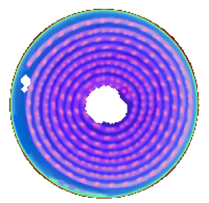
The result is colorized images with what the team calls revolutionary image clarity and a better understanding of an object’s composition.
“We get a more accurate representation of the shape and definition of that object, which is going to allow us to make unprecedented measurements and unprecedented observations,” Jimenez said.
Far-reaching applications
The team sees this as a major advancement for X-ray technology with a wide range of uses, from airport security and quality control to nondestructive testing and advanced manufacturing.
They also hope its impact will improve medical diagnostics.
“With this technology, you can see even slight differences between materials,” Jimenez said. “We hope this will help better identify things like cancer and more effectively analyze tumor cells. In mammography you are trying to catch something before it grows. In breast tissue, it’s hard to identify the different dots, but with colorization you have a sharper beam and higher resolution image that increases the system’s capability to detect a microcalcification. It’s really exciting to be a part of that.”
“From here we will continue to innovate,” Collins said. “We hope to identify threats faster, diagnose diseases quicker and hopefully create a safer, healthier world.”
For additional video, click here.
The team was recently awarded an R&D 100 award for their technology. They were among six winners from Sandia. Click for R&D 100 Submission video with soundbites.