ALBUQUERQUE, N.M. — A Sandia National Laboratories research team led by Anup Singh is taking a new approach to studying how immune cells respond to pathogens in the first few minutes and hours of exposure.
Their method looks at cells one at a time as they start trying to fight the invading pathogens.
Called the Microscale Immune Studies Laboratory (MISL) Grand Challenge, the work is in its second of three years of funding by the internal Laboratory Directed Research and Development (LDRD) program. Sandia is partnering on the project with the University of Texas Medical Branch (UTMB) at Galveston and the University of California, San Francisco (UCSF).
Sandia is a National Nuclear Security Administration (NNSA) laboratory.
Singh says the researchers are interested in studying the early events in immune response when a pathogen invades a body. Understanding the early steps could lead to better ways to diagnose and stop disease before there are symptoms and development of more effective therapeutics.
Most existing research into how immune cells respond has been done by looking at large cell populations. The Sandia researchers say information gathered from a large population of cells may mask underlying mechanisms at the individual cell level.
“Cells have different life cycles, just like any living being. And not all cells are exposed to the pathogen at the same time,“ Singh says. “We wanted to look at cells in the same life cycle and same infectious state. This can only be done cell by cell. We also want to study populations, but one cell at a time.”
The research is possible because of advances in several Sandia-developed tools, including:
- Microfluidics that allows researchers to do single-cell experiments
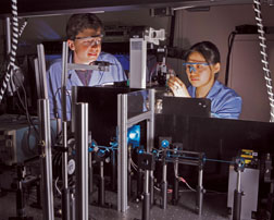
- Advanced imaging that allows researchers to image individual cells with much higher information content than possible with current commercial imaging technologies
- Powerful computational modeling that allows researchers to make sense of data obtained from microfluidic analysis and imaging

Real immune cells are short-lived outside of bodies. To do the type of experiments they wanted, the researchers needed cells that can stay alive more than a couple of hours, have the ability grow and represent a relevant model of human immune cells. They obtained “immortalized mouse immune cells” from a collaborator at UCSF that have the needed life span, and are accepted as a model system by the immunology research community.
“We’re starting with robust and well-characterized cells, which really simplifies development of our new technologies and methods,” Singh says. “We’ll soon be working with other cell types, though, like white blood cells directly isolated from human patients. Our approach is designed to be flexible enough to handle many different cell types, and it also minimizes the number of cells needed for analysis, so it should enable us to do some unique studies on rare cell types.”
Proteins in the cells of interest are tagged with fluorescent molecules, essentially colored dyes. The dyes range from green to red and give researchers the opportunity to track proteins and see, for example, the dynamic cellular production of proteins or protein-binding processes inside or on the surface of the cells.
The team is developing one platform with two complementary microfluidic modules — one for trapping and imaging viable cells during stimulation with pathogens. The other combines cell preparation steps, cell selection and sorting followed by analysis of protein content in the selected cell subpopulations.
“In effect, we are taking many work-horse technologies such as confocal microscopy, flow cytometry and immunoassays and combining them into one compact, miniaturized platform using our unique microfluidic and imaging tools,” Singh says.
Hyperspectral fluorescence imaging with multivariate curve resolution (MCR) is used to image the tagged proteins and provide quantitative measurements on multiple proteins simultaneously. The goal is to analyze as many as 10 to 40 proteins and cellular stains at a time in three dimensions.
The end results of the imaging and protein analysis are large amounts of data that must be categorized and understood. Computational modeling is then used to develop network models from experimental data and predictive modeling generates hypotheses to be tested next.
Singh says using an integrated microfluidic platform sets Sandia apart from the rest of the world. Sandia researchers have been working in the area of microfluidics — the science of designing, manufacturing, and formulating devices and processes that deal with volumes of fluid on the order of nanoliters — since the 1990s and have a good understanding about how to use microfluids to analyze cell activity. The microfluidic platform is fast and highly parallel and can perform hundreds of measurements 50 to 100 times faster than alternate methods.
Singh says the end goal is to make a benchtop miniaturized system expected in about two years. It would be placed in Biosafety Level 3 or 4 labs to study immune response to highly pathogenic organisms. He notes the integrated platform, biological reagents and computational models developed under this project have applicability beyond infectious disease research. These technologies can also be used for studying cellular signaling involved in diseases such as cancer or by pharmaceutical companies for biomarker discovery.
More information can be obtained at http://roswell.ca.sandia.gov/anup.

