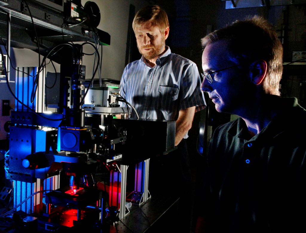
Sandia National Laboratories researchers Michael Sinclair (foreground) and David Haaland prepare a hyperspectral confocal microscope for measurement of a biological specimen. The new microscope, shown on the left, was designed and fabricated at Sandia. (Photo by Randy Montoya)
ALBUQUERQUE, N.M. —Sandia National Laboratories will demonstrate a new hyperspectral confocal fluorescence microscope Friday, Aug. 8 from 9 a.m.-2 p.m. MDT in Bldg. 897 on Kirtland Air Force Base. This patent-protected and patent-pending technology has been combined with Sandia’s unique and proprietary multivariate algorithms and software to form a complete system for the extraction of quantitative image information from hyperspectral images.
The microscope has been used for multiplexed live-cell imaging at diffraction-limited spatial resolutions in a variety of biological applications. Included in the hyperspectral imaging system is spectral image viewing software to view both the raw image data as well as the results from the multivariate curve resolution (MCR) analyses, says Dave Haaland, a researcher in Sandia’s Biomolecular Analysis and Imaging Department.
The hyperspectral microscope uses laser excitation and collects 512 spectral emission wavelengths at each voxel in the image over the spectral range from 500 to 800 nanometers at a spectral resolution of 1-3 nm and at an imaging rate of 8,300 spectra/second (with extension to 64,000 spectra/sec in the future).
Sandia’s proprietary MCR software allows very rapid “discovery” of all emitting fluorescence species in the image and determination of their relative concentrations throughout the image without any a priori information. With this new system, many fluorophores can be monitored simultaneously without cross talk to achieve higher throughput, greater quantitative accuracy, and increased reliability.
“Sandia is seeking to commercialize this technology by partnering with interested firms through negotiated licensing agreements,” says Brent Burdick, licensing executive in Sandia’s Licensing and Intellectual Property Management Department. “Accordingly, the Aug. 8 demonstration is being held to allow firms interested in commercializing the technology an opportunity to see first-hand how the microscope works.”
Due to processing requirements, registrations must be received by Friday, July 25. For further information on the microscope or attending the demonstration, contact Jonathan Gardner at jgardn@sandia.gov or (505) 845-7653.

